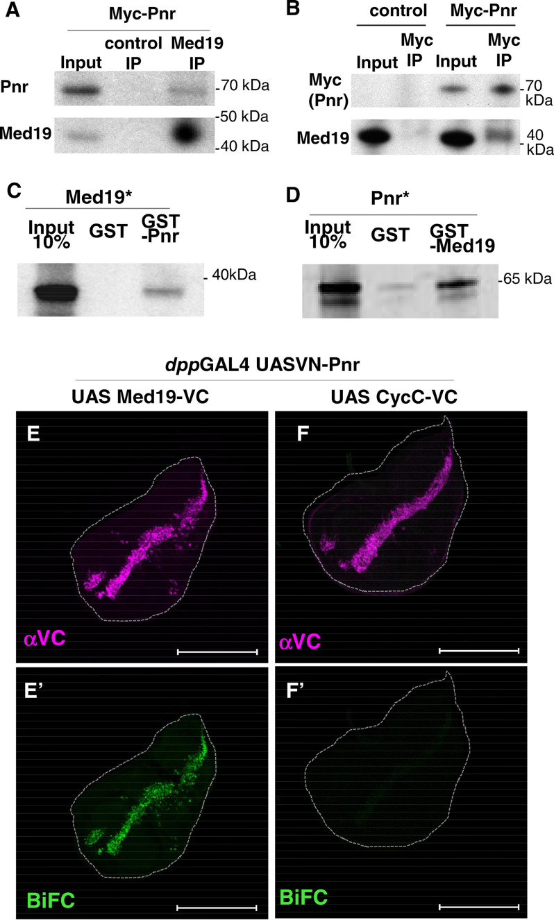
Figure 2.
Med19 physically interacts with GATA/Pnr in cellulo, in vitro, and in vivo. A and B, co-IP experiments from S2 cells transfected with pAct-Myc-Pnr using anti Med19 antibody or preimmune serum (control IP) (A). The reverse experiment (B) was performed using anti-Myc beads from control cells or cells transfected with pAct-Myc-Pnr. Western blot assays using αMed19, αPnr, or αMyc antibodies are shown. C and D, autoradiographs from GST pulldown assays between GST-Pnr and 35S-labeled (asterisk) in vitro–translated Med19 (C), and between GST-Med19 and in vitro–translated 35S-Pnr (D). E–F′, BiFC assays using VN-Pnr and either Med19-VC (E) or CycC-VC (F). Expression of fusion proteins along the A/P boundary of the wing disc is under the control of dppGAL4 driver. Immunostaining shows expression of VC constructs (magenta, E and F), and BiFC signals are shown in green (E′ and F′). Scale bars, 200 μm.

