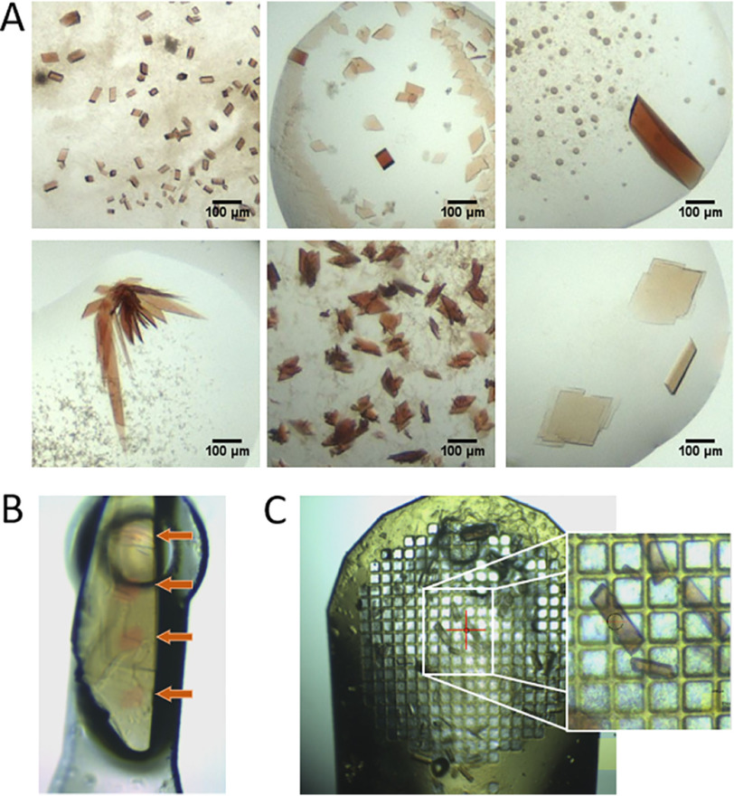
Figure 2.
Representative crystal forms and sizes of the investigated heme proteins. A, crystal forms and sizes of KpDyP (top, left), NdCld (top, middle), CCld (top, right), hhMb (bottom, left), AvTsdA (bottom, middle), and LmChdC (bottom, right). B, visualized impact of X-ray–induced reduction as seen on a mounted crystal of LmChdC. Red dots (indicated by the orange arrows) show areas hit by the X-ray beam. C, 750-μm mesh loaded with crystals from KpDyP and close up. Mesh cryo-cooling allows the rapid manual collection of up to 30–50 crystals without the need for time-consuming sample changing.

