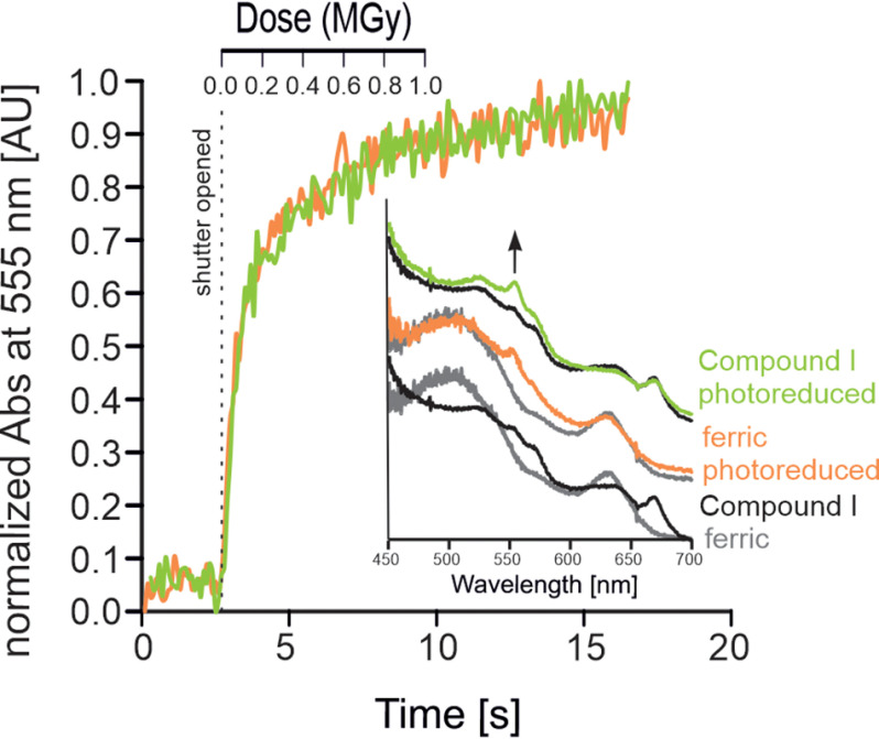
Figure 5.
Kinetics of radiation-mediated reduction of Compound I to Fe(II) in KpDyP. Shown are time- and dose-dependent changes in absorption at 555 nm of the ferric resting state (orange) and the Compound I intermediate (green) of KpDyP upon photoreduction (10% transmission, flux = 4.2 × 10¹¹ photons s−1). Inset (bottom), overlay of single-crystal UV-visible spectra of the ferric (gray) resting state and the Compound I intermediate (black) obtained by soaking with H2O2. Inset (middle), ferric resting state before (gray) and after (orange) exposure. Inset (top), Compound I intermediate before (black) and after (green) exposure.

