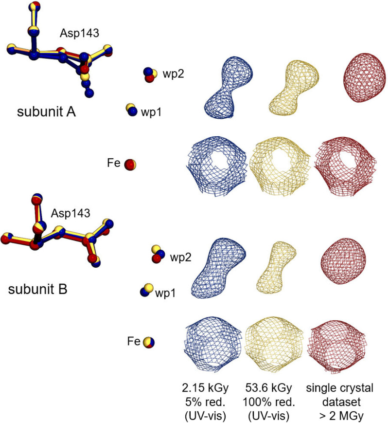
Figure 6.
Structural changes upon reduction of ferric high-spin KpDyP. Asp-143 (sticks), distal active-site water molecules, and the heme iron (sphere) are represented from KpDyP subunit A (top) and B (bottom) for the 5% reduced (blue, 6RQY), the 100% reduced (gold, 6RR8) structure, based on UV-visible spectroscopy, from the multicrystal approach and for the completely reduced routine single-crystal data set (red, 6FKS). 2mFo − DFc (contoured at 1 σ) electron density maps for the represented molecules are shown in the respective colors on the right.

