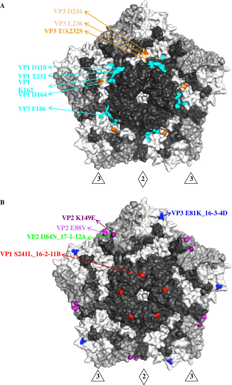
Fig 3. Antigenic determinants on the viral capsid of genotype C1 EV-A71.
(A) Canyon epitopes, including VP1 D110, VP1 T232, VP1 K162, VP1 D164 and VP3 F186, recognized by human anti-EV-A71 MAbs 16-2-9D, 16-2-12D, 16-3-3C and 16-2-2D, were colored in cyan [24]. The surface residue 232 of VP3, located at the canyon region of capsid, was distinct in genotype C1 EV-A71 (S2 Fig) and interacted with VP3 D234 and L236 canyon residues (colored in orange). (B) Epitope mapping on the viral capsid by genotype C1 EV-A71-neutralizing MAbs. Escape variants were selected using MAbs 16-2-11B, 16-3-4D and 17-1-12A and revealed single amino acid substitutions at capsid residues VP1 S241 (colored in red), VP3 E81 (colored in blue) and VP2 D84 (colored in green), respectively. VP2 E88V and VP2 K149E were previously identified in the escape variant of genotype B5 and C4 EV-A71 with MAb 17-1-12A [24]. The surface view of pentamer shown with the 5-fold vertex at the center are created using the software program PyMOL (PDB 3VBS). The capsid VP1 protein is colored in black, VP2 colored in grey and VP3 colored in white. Abbreviations: 3, 3-fold axis; 2, 2-fold axis.

