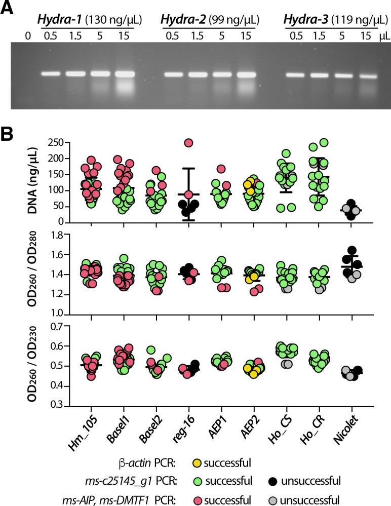
Fig 2. Direct genomic DNA amplification from single Hydra polyp.

(A) PCR amplification of β-actin genomic DNA from three 4–6 mm long, non-budding AEP animals dissociated and resuspended each in 50 μl water. Various amounts (from 0 to 15μL) of the resulting macerates were used as PCR template for β-actin amplification to estimate PCR efficiency. (B) Graphic representation of DNA concentration and DNA purity as deduced from OD measurements at 260, 230 and 280 nm wave lengths. Each dot represents either the OD260 value or the 260/280 or 260/230 OD value ratios obtained from a single polyp. For each DNA, the efficiency of PCR amplification is indicated with a color code. Note the lower DNA content in most reg-16 polyps that were fixed in PFA and stored in methanol for months prior to rehydration, maceration and DNA amplification.
