FIGURE 2:
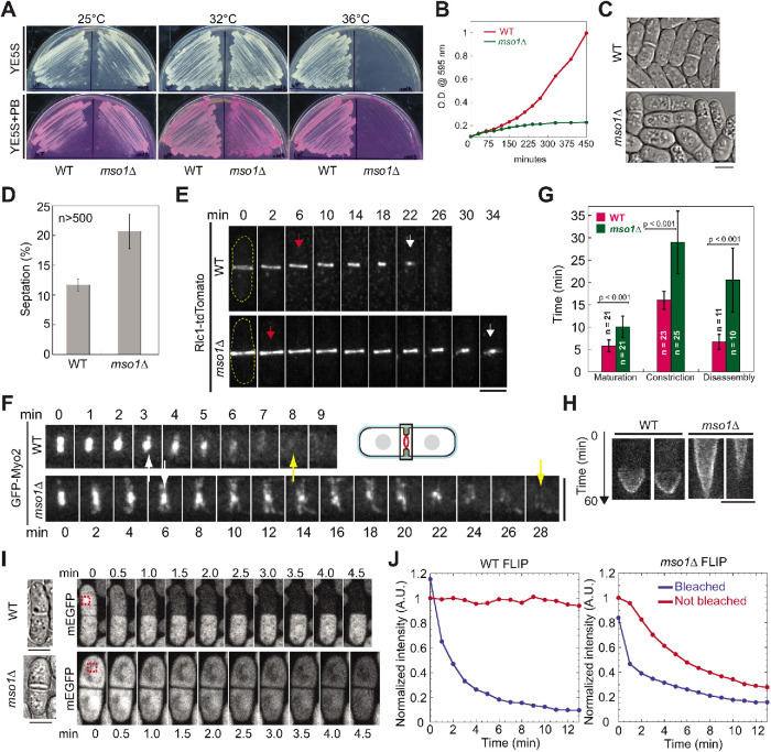
Mso1 is important for cytokinesis. (A) Wild-type (WT) and mso1∆ cells grown on YE5S and YE5S + PB plates at 25°C, 32°C, and 36°C. (B) Growth curves of WT and mso1∆ cells in liquid cultures at 36°C. (C, D) DIC images (C) and septation index (D; mean ± SD) of WT and mso1∆ cells grown in YE5S liquid media at 36°C for 4 h. (D) For each strain, three sets of data were measured, with all sets having >500 cells. (E–G) The contractile ring (marked with Rlc1 or Myo2) is defective in mso1∆ cells after growth at 36°C for 2 h. (E, F) Time course of contractile-ring constriction (E) and disassembly (F) in WT and mso1∆ cells. The arrows indicate the start (red) and end (white) of ring constriction and disappearance (yellow) of Myo2 signal. (G) Quantification of ring maturation (Rlc1), constriction (Rlc1), and disassembly (Myo2). See Materials and Methods for the definition of each stage. Error bars are SDs. (H) Kymographs of different Ain1-mEGFP expressing cells showing ring constriction in WT and mso1Δ cells. Kymographs are 60 min from top to bottom with each line representing 1 min. (I, J) Exchange of free GFP between daughter cells is observed in mso1∆ but not in WT cells after ring constriction. Time courses (I) and intensity measurements (J) of fluorescence loss in photobleaching (FLIP) assays in WT and mso1∆ cells after growth at 36°C for 2 h. The box with red dash lines in I marks the bleached region. Bars, 5 µm.

