FIGURE 1:
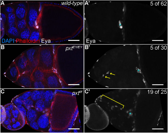
Prostaglandins regulate border cell cluster integrity. (A–C′) Maximum projections of three confocal slices of S10 follicles of the indicated genotypes; anterior is to the left. (A–A′) wild type (yw). (B–B′) pxtEY03052/EY03052 (pxtEY/EY). (C–C′) pxtf01000/f01000 (pxtf/f). (A–C) Merged images: Eyes absent (Eya), white; phalloidin (F-actin), red; and DAPI (DNA), blue. (A′–C′) Eya, white; images were brightened by 50% in Photoshop for better visualization. The nuclei of the border, stretch follicle, and centripetal cells are marked by Eya staining; polar cells are not marked. By S10, the intact border cell cluster is normally located at the nurse cell/oocyte boundary (A–A′, cyan asterisk). In pxt mutants, despite the majority of the cluster reaching the boundary (cyan asterisk), cells are often left behind along the migration pathway (B–C′); the frequency of S10 follicles exhibiting trailing border cells is indicated in the top right of panels A′–C′. These cells can exist as single cells or pairs of cells being left behind (B–B′, yellow arrows), or long continuous chains of cells being left behind (C–C′, yellow bracket). Scale bars = 50 μm.

