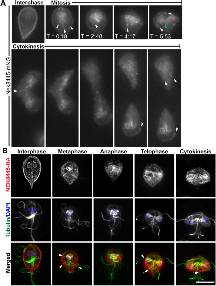
FIGURE 3:
Nek8445 relocalizes during mitosis to associate with axoneme microtubules. (A) Still images from a time-lapse movie of Nek8445-mNG showing Nek8445 translocating from the cell cortex to the nuclear envelope, and axoneme microtubules (see Supplemental Video S2). White arrowheads indicate Nek8445 in association with cytoplasmic axonemes; cyan arrowhead marks nascent axonemes pointing into the developing furrow. (B) Immunofluorescence analysis of Nek8445-HA (red), microtubules (6-11B-1 antibody; green), and nuclei (DAPI; blue) throughout the cell cycle. Scale bar = 5 µm.

