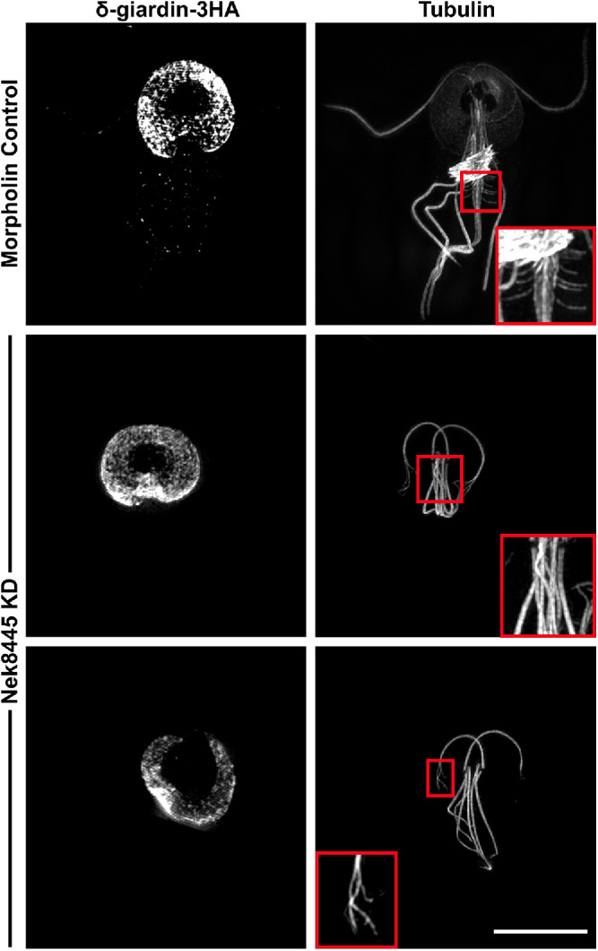
FIGURE 8:

Structured Illumination superresolution imaging of δ-giardin-3HA and tubulin (6-11B-1 antibody) in morpholino-treated cells. Nek8445-depleted cells lack median bodies, a funis, and the axonemes have a fraying defect. Control inset shows a magnified view of funis microtubules. Middle inset shows that the Nek8445-depleted cells lack both a median body and funis. The bottom image shows an example of a Nek8445-depleted cell with abnormal disk morphology and splayed axoneme tips. Scale bar = 5 µm.
