FIGURE 1:
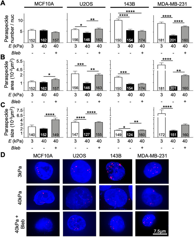
Paraspeckle expression on 3 kPa and 40 kPa in MCF10A, U2OS, 143B, and MDA-MB-231 cells. (A) The average number of paraspeckles per nucleus was higher in cancer cell lines cultured on 3 kPa hydrogels compared with 40 kPa hydrogels in U2OS (5.75 vs. 4.28), 143B (9.88 vs. 5.47), and MDA-MB-231 (9.22 vs. 3.44) cell lines. No difference in paraspeckle number was observed in the MCF10A cell line. Treatment of cells cultured on 40 kPa hydrogels with blebbistatin revealed an increase in paraspeckle number in the U2OS (4.28 to 6.00) and MDA-MB-231 (3.44 to 7.65) cell lines. (B) Paraspeckle total area showed the same trend in U2OS (0.021 µm2 vs. 0.0094 µm2), 143B (0.017 µm2 vs. 0.0082 µm2), and MDA-MB-231 (0.055 µm2 to 0.0054 µm2) cell lines but not in the MCF10A cell line. Blebbistatin treatment also resulted in increased paraspeckle area in MCF10A, 143B, and MDA-MB-231 cell lines. (C) Analysis of paraspeckle size revealed that paraspeckles appeared larger in size in cancer cells cultured on 3 kPa hydrogels vs. 40 kPa hydrogels in U2OS (0.0034 µm2 vs. 0.0019 µm2), 143B (0.0016 µm2 vs. 0.0014 µm2), and MDA-MB-231 (0.0069 µm2 vs. 0.0015 µm2) cell lines. Blebbistatin treatment further increased paraspeckle size in all cell lines. (D) Representative images showing paraspeckles (red) in nuclei (blue) taken at 60× magnification. Scale bar = 7.5 µm. Data are shown as mean ± SEM. Numbers of nuclei used in analyses were indicated per bar graph. *, p < 0.05; **, p < 0.01; ***, p < 0.001, and ****, p < 0.0001.

