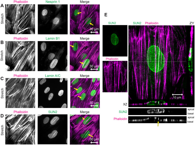
FIGURE 4:
Stretch-stimulated LINC components include Nesprin, SUN, and Lamin proteins. (A–D) Widefield fluorescence microscopy of F-actin (phalloidin, magenta) and LINC components (green) including Nesprin 1, Lamin B1, Lamin A/C, and SUN2 in stretch-stimulated cells. Merged images show codistribution of actin and LINC proteins (yellow arrows) in nuclei aligned perpendicular to the stretch vector (indicated by a horizontal double-headed arrow of 30 micron scale). (E) Confocal microscopy of stretch-stimulated fibroblast stained with SUN2-specific antibody (green, Burke #3.1E mab) and Phalloidin (magenta), shown as maximum intensity projections with 10 micron scale bar. Orthogonal z sections are designated by dashed lines in the x (below merge) and y (right of merge) planes. Grayscale z sections are shown with apical surface on top and basal surface on bottom. SUN2 is distributed throughout the NE and F-actin is distributed across the top of nucleus and along the flat cell edges. SUN2 localizes with one prominent actin SF (yellow arrow) but not with a neighboring SF crossing the nucleus; see associated Supplemental Videos S1–S3.

