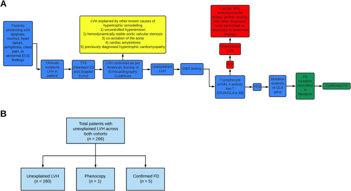
Fig 1.
(A) Schematic showing overall design of study and flow of patients across both sites. (B) Flow diagram depicting breakdown of patients across both Edmonton and Hong Kong cohorts. LVH indicates left ventricular hypertrophy; TTE, transthoracic echocardiography; DBS, dried blood spot; a-GAL A, α-galactosidase A enzyme; IDUA, alpha-L-iduronidase; GLA, α-galactosidase gene; FD, Fabry Disease; HCQ, hydroxychloroquine.

