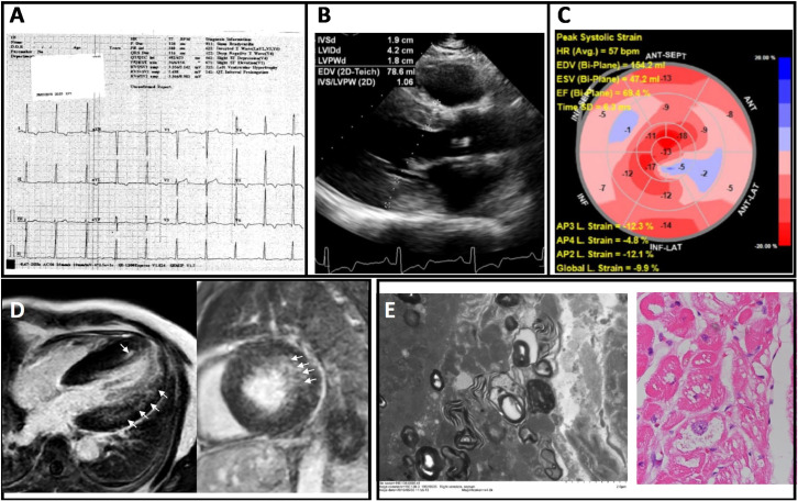
Fig 4. Electrocardiographic, imaging, and endomyocardial biopsy findings of a 60 year-old man originally diagnosed with nonobstructive hypertrophic cardiomyopathy.
(A) Electrocardiography showed high voltage in limb and precordial leads consistent with LVH; (B) Transthoracic echocardiography showed LV walls thickening with maximum wall thickness of the septum and posterior wall measuring 1.7 cm and 1.9 cm respectively, with LV ejection fraction = 64%; (C) Global longitudinal strain was reduced (–9.9%); (D) Cardiac MRI showing long-axis (left) and short-axis (right) views illustrating concentric LVH and mild patchy late gadolinium enhancement in the lateral wall and the apical septum (arrows). (E) Pathological findings based on electron microscopy of endomyocardial biopsy showing central vacuolations and lamella bodies in cardiomyocytes (left) and PAS staining showing vacuolation and disarray (right), consistent with Fabry cardiomyopathy.

