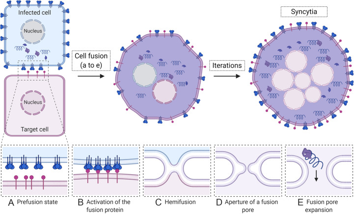
Fig. 2.
Syncytia formation. Infected cells (blue) display a high concentration of the viral fusion protein (shown as blue spikes) on cell surfaces. When infected cells come into proximity with an uninfected cell (purple), several sequential steps lead to syncytia formation and spread of infection. (A) Initially, the fusion protein is in an inactive prefusion state; (B) upon the proper stimulus (such as binding a receptor shown in purple on the target cell), the fusion protein is triggered, which induces conformational changes, insertion of a fusion peptide into the adjacent membrane, and conformational changes that bring both membranes, viral and cellular into close proximity, followed by a partial merging known as hemifusion (C). Full merger of both membranes results in the opening of a fusion pore (D) that expands allowing the mixing of cytoplasmic material and spread of viral components between cells (E). Iterations of the fusion process result in formation of large multinucleated syncytia.

