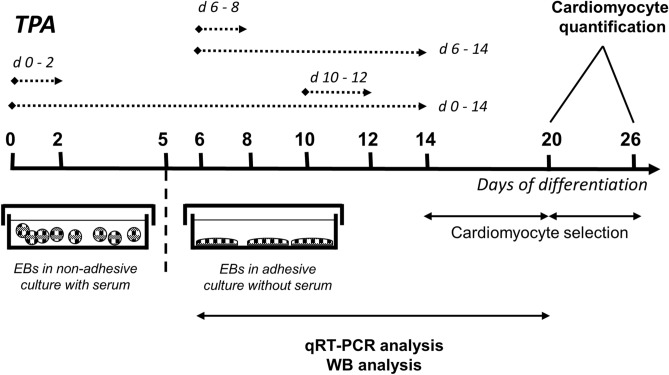
Figure 1.
Experimental setup employed for comparison of the cardiomyogenic differentiation of ES cells exposed to different TPA treatment. Cells were treated with 1 µM of TPA on Days 0–2 (d 0–2), Days 0–14 (d 0–14), Days 6–8 (d 6–8), Days 6–14 (d 6–14), and Days 10–12 (d 10–12) of differentiation. The number of cardiomyocytes was calculated on Days 20 and 26. Additionally, cardiomyocyte markers were analysed between Day 7 and Day 20 by qRT-PCR and Western blot method.

