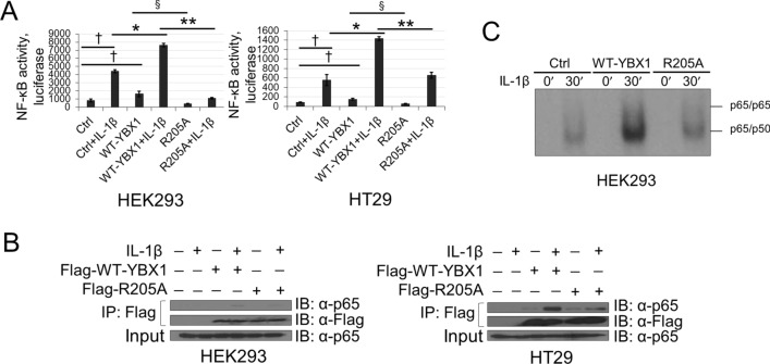
Figure 4.
R205A attenuates YBX1-mediated NF-κB activation, p65 DNA binding and complex between p65 and YBX1. (A) NF-κB luciferase assay performed in HEK293 (left) and HT29 (right) cells stably expressing the WT-YBX1 or R205A construct with or without 10 ng/ml IL-1β stimulation. Luciferase readings were normalized to both protein concentration and internal control β-galactosidase. (B) Co-immunoprecipitation of YBX1 and endogenous p65. HEK293 and HT29 cells stably overexpressing Flag-WT-YBX1 or Flag-R205A were treated with 10 ng/mL of IL-1β for 1 h or were left untreated. YBX1 primarily complexes with p65 under IL-1β stimulation. Anti-p65 and Anti-Flag IP images were obtained from the same blots that were stripped and reprobed for total Flag but developed at different exposures. Input images were obtained from different blots and developed at different exposures. (C) EMSA to determine DNA binding ability of p65 performed with extracts from HEK293 cells overexpressing WT-YBX1 or R205A. Cells were left untreated or stimulated with 10 ng/ml IL-1β for 30mins. The data represent the means ± SD from three independent experiments. †p < 0.05 vs. Control (Ctrl) group; *p < 0.05 vs. Ctrl + IL-1β group; §p < 0.05 vs. WT-YBX1 group; **p < 0.05 vs. WT-YBX1 + IL-1β group.

