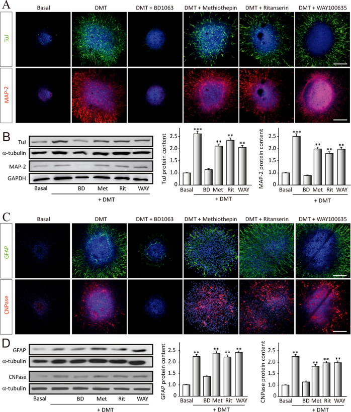
Fig. 2. N,N-dimethyltryptamine (DMT) promotes stem cell differentiation toward all neural phenotypes.
SGZ-derived neurospheres were cultured for 7 days in the presence of DMT alone or in combination with the antagonists BD1063 (BD), methiothepin (Met), ritanserin (Rit), and WAY100635 (WAY) and then were adhered on a substrate and allowed to differentiate for 3 days. a Confocal fluorescent images showing the expression of the neuronal markers β-III-Tubulin (TuJ-1 clone, green) and MAP-2 (red) in NS (n = 8 per condition). DAPI was used for nuclear staining. Scale bar = 50 μm. b Representative western blots and quantification of β-tubulin and MAP-2. c Immunofluorescence images showing NS expressing the glial fibrillary acidic protein (GFAP, red) that stains astrocytes, and in green, the oligodendrocyte marker CNPase (n = 8 per condition). DAPI was used for nuclear staining. Scale bar = 50 μm. d Representative western blots of CNPase and GFAP and quantification. Results are the mean ± SD of four different cellular pools with three independent experiments/pool (n = 12). After confirming the significance of the primary findings using ANOVA, a significance level of P < 0.05 was applied to all remaining post hoc (Tukey test) statistical analyses. **P ≤ 0.01; ***P ≤ 0.001 indicate significant results versus non-treated (basal) cultures.

