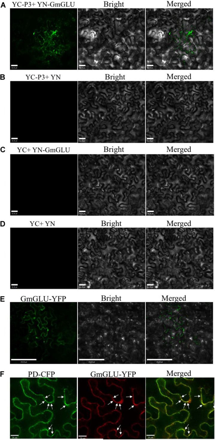
FIGURE 2.
Bimolecular fluorescence complementation assays demonstrating SMV P3 and GmGLU interaction in cytomembrane and epidermal cells of N. benthamiana. (A) SMV P3 and GmGLU were fused to N- and C-terminal of yellow fluorescent protein (YFP) respectively. YFP fluorescence in N. benthamiana leaves agroinfiltrated with pEarleyGate202-YN-GmGLU+pEarleyGate201-YC-P3. (B–D) Negative controls in the BiFC assay of GmGLU and SMV P3 interaction. Bar indicates 36 μm. (E) Fluorescence of YFP fused to GmGLU protein of host in N. benthamiana leaves under spectral confocal laser microscope. Bar indicates 140 μm. (F) Co-expression of plasmodesmata (PD) marker fused with cyan fluorescent protein (CFP) and GmGLU protein fused with YFP. GmGLU protein accumulated in dots on cytomembrane (PD marker sites). Bar indicates 20 μm.

