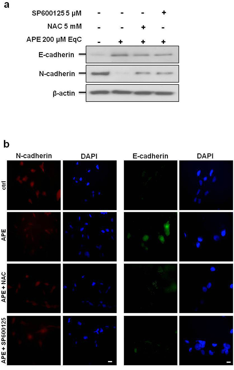
Figure 4.
ROS/JNK signaling activation mediated APE-induced switch from N- to E-cadherin expression. (a) The levels of E-cadherin and N-cadherin in MDA-MB-231cells treated with APE 200 μM EqC for 48 h with or without 1 h pretreatment with 5 mM NAC and/or 5 μM SP600125 were measured by Western blot. β-actin was used as loading control. Results were obtained from at least three independent experiments. The full-length blots are included in Supplementary Information (Fig. S5). (b) Representative immunofluorescence images of N-cadherin and E-cadherin in MDA-MB-231 cells treated or not (ctrl) with 200 μM EqC APE for 24 h, with or without 1 h pretreatment with 5 mM NAC and/or 5 μM SP600125 are shown. Nuclei were stained with DAPI (blue), while anti N-cadherin (red) or E-cadherin (green) antibodies were used to detect the corresponding proteins. Images are representative of three independent experiments. Scale bar, 5 μm.

