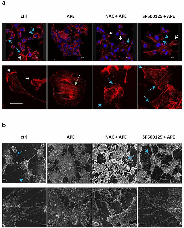
Figure 6.
ROS/JNK signaling activation mediated APE-induced changes in podia formation, actin cytoskeleton remodeling, and cell morphology. (a) Representative confocal images of F-actin architecture in MDA-MB-231 cells treated or not (ctrl) with APE 200 µM EqC for 24 h with or without 1 h pretreatment with 5 mM NAC and/or 5 μM SP600125. Cells were cultured on fibronectin-coated coverslips and stained with Alexa Fluor 546 Phalloidin. Lamellipodia (white arrows), stress fibers (green arrows), and filopodia (blue arrows) were identified under a confocal microscope. Scale bar: top row, 37.5 μm; bottom row, 20 μm. (b) SEM images of MDA-MB-231 cells treated or not (ctrl) with APE 200 µM EqC for 24 h with or without 1 h pretreatment with 5 mM NAC and/or 5 μM SP600125. Control and pretreated cells showed typical mesenchymal characteristics such as elongated spindle-like shape with cytoplasmic protrusions like filopodia (blue arrow) and few globular rounded cells. APE treated cells showed a wide cytoplasm, tight cell–cell contacts, and an increase of flattened very large cuboid cells which appeared more grouped, resembling epithelial-like sheets. Scale bar: top row, 10 μm; bottom row, 1 and 2 μm.

