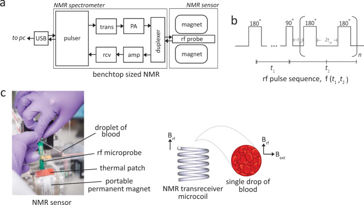
Fig. 1. Two-dimensional NMR T1-T2 correlational spectroscopy for molecular phenotyping of blood.
a Schematic diagram of the bench-top sized NMR-based POCT system. The applied radio frequencies were centered at 21.57 MHz, which corresponds to the Larmor frequency of water-proton in 0.5 Tesla of the permanent magnet. The 90-degree pulse used is 10 μs. The whole system is lightweight (<2 kg) and portable suitable for in situ measurements. The abbreviations are; USB Universal Serial Bus, trans Transmitter, rcv Receiver, amp pre-amplifier, PA power amplifier, rf radio-frequency, and PC personal computer. b The pulse sequence used for the T1-T2 correlational spectroscopy is the modified inversion recovery with CPMG observation. It is encoded for a period of t1 and subsequently spaced for a period of t2 for n-train pulses, in entirely in analogous to the two-dimensional NMR spectroscopy in the frequency domain. The relaxation properties can be used as a highly sensitive and specific molecular probe, and provide important molecular motion (e.g., correlational relaxation, diffusion properties), which is not readily available in NMR spectra in the frequency domain. c A single drop of whole blood contained in a microcapillary tube was spun using standard hematocrit centrifuge (6000 × g, 1 min) to separate and concentrate the RBCs from the plasma. The capillary tube is then loaded into a permanent magnet. The tube was adjusted as such that the radio frequency coil (inner diameter of 1.20 mm) focuses on the packed RBCs (enrichment part). This is essential to have ‘clean signal' from the RBCs without (or with minimal) interference of blood plasma.

