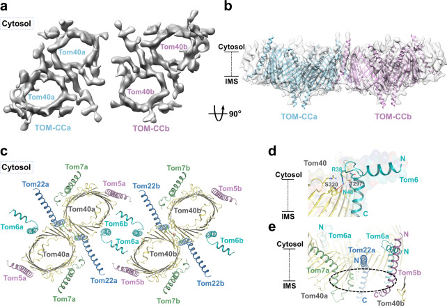
Fig. 4. Structure of the tetrameric TOM complex.
a A 8.5-Å-resolution cryo-EM reconstruction of the tetrameric TOM complex. Two dimeric units are indicated by a and b. Cytosol views are shown. b Dimeric TOM complex models fitted in the map. Two dimeric units are indicated by a and b. Side views are shown. c Fitted model of tetrameric TOM complex. Cytosol views are shown. d Interactions between Tom6F34–R42 and Tom40. The polar interactions are indicated by red dotted lines. Side views are shown. e A potential binding pocket formed by Tom22, Tom7 from one dimer and Tom5 from the other dimer. The potential pocket is indicated with dotted cycles. Side views are shown.

