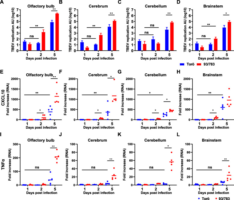
Fig. 2.
Viral burdens in brains after intracranial infection with 93/783 and Torö. Six- to 10-week-old mice were infected intracranially with 102 FFU of 93/783 or Torö. Viral burdens in the olfactory bulb (a), cerebrum (b), cerebellum (c), and brainstem (d) were determined by qPCR; expression levels were normalized to the endogenous GAPDH expression and calculated using the ΔΔCT method. CXCL10 and TNFα expression levels were detected with qPCR in the olfactory bulb (e, i), cerebrum (f, j), cerebellum (g, h), and brainstem (h, l), normalized to GAPDH and expression levels in Torö day 1 for the different brain regions (N = 3 − 5). Asterisks indicate statistical significance calculated using Mann-Whitney test (ns = not significant, *P < 0.05, **P < 0.01)

