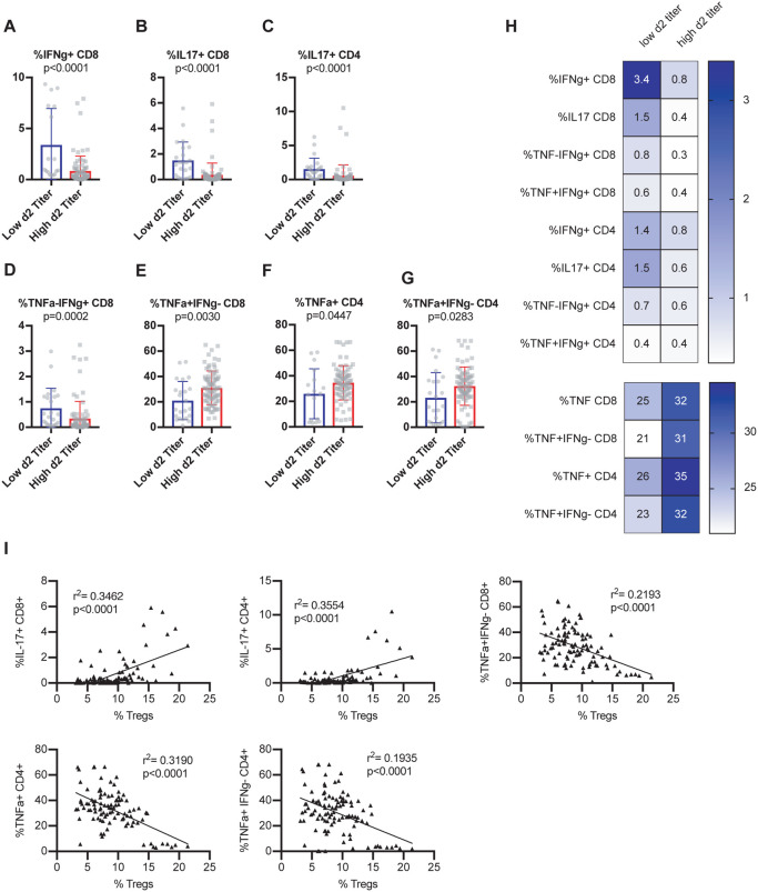
Figure 3. Early viral control upon infection correlates with baseline T cells with a potential to express IFNg or IL17 rather than TNF.
Age-matched female CC-RIX were infected intranasally with SARS-CoV MA15 and lung viral loads at day 2 post-infection were used to select CC-RIX lines with extreme phenotypes: “Low 2d Titer” or “High 2d Titer”, as indicated in Figure 1. Mice from a second cohort of 3–6 age-matched male mice of these selected lines were euthanized and splenic cells were treated with anti-CD3/CD28 for intracellular cytokine staining assessment of (A) %IFNg+ of CD8 T cells, (B) %IL-17+ of CD8 T cells, (C) %IL-17+ of CD4 T cells, (D) %TNF-IFNg+ of CD8 T cells, (E) %TNF+IFNg− of CD8 T cells, (F) %TNF+ of CD4 T cells, and (G) %TNF+IFNg− of CD4 T cells. Statistical significance was determined by Mann-Whitney test. (H) Heat maps were made to compare the average percent of the indicated cell populations. No statistical significance (p>0.05 by Mann-Whitney test) was found for any comparisons except those indicated in Figures 3A–G. (I) The correlation between the baseline splenic frequency of Tregs (% Foxp3+ of CD4 T cells) and % of CD8 T cells that are IL-17+, % of CD4 T cells that are IL-17+, % of CD8 T cells that are TNF+IFNg−, % of CD4 T cells that are TNF+, and the % of CD4 T cells that are TNF+IFNg− are shown with linear regressions for mice from all CC-RIX lines with low or high day 2 titer.

