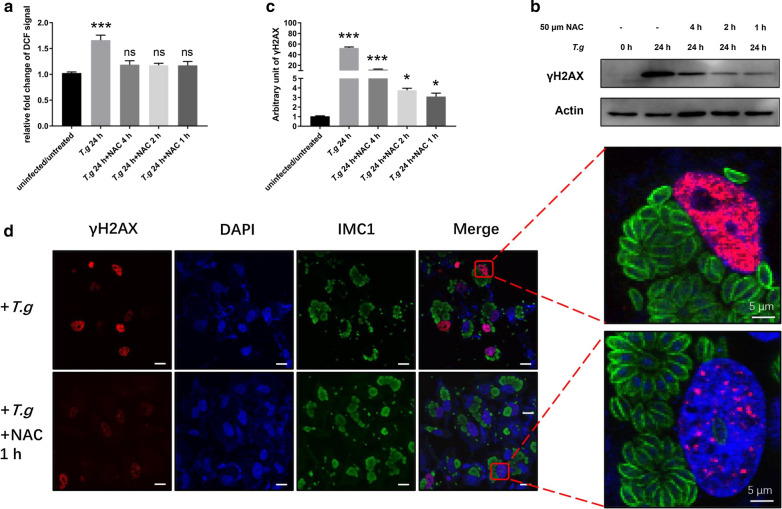
Fig. 3.
Reactive oxygen species contributed to DNA damage. HeLa cells were seeded in a 24-well plate and infected with T. gondii RHΔku80 at a MOI of 10:1 for 24 h. N-acetylcysteine (NAC) at 50 μm was then applied to the infected cells for 1, 2 or 4 h. Antibodies to γH2AX, actin, and IMC serum were all used at a 1:1000 dilution for the western blot and at a 1:200 dilution for the immunofluorescence assay (IFA). a NAC treated T. gondii-infected cells were stained with 10 μm H2DCFDA and fluorescence signals were recorded. Data are presented as the mean ± SD (n = 3). b γH2AX was detected by western blot. c Quantification of γH2AX shown in b. Mean ± SD (n = 3) of γH2AX levels are shown with that of uninfected cells set as one arbitrary unit. d γH2AX assayed by IFA. Key: red, γH2AX; green, IMC1; blue, DAPI. All experiments were performed in triplicate. *P ≤ 0.05, ***P ≤ 0.001; ns: not significant. Scale-bars: 30 μm

