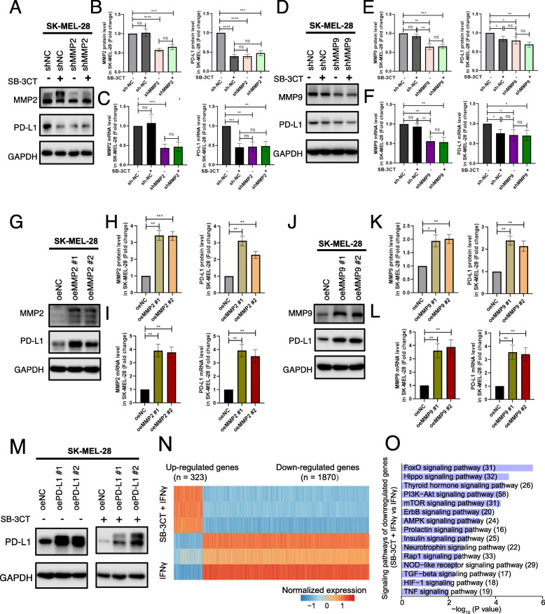
Fig. 5.
Regulation of PD-L1 expression through MMP2/9 has an anti-tumor effect. a–f Analysis of PD-L1 expression of SK-MEL-28 melanoma cell line with overexpression (oe) MMP2 (a–c), oeMMP9 (d–f). Western blot (a, d), quantification (b, e), and RT-PCR analysis (c, f) of MMP2, MMP9, and PD-L1 protein or mRNA expression. g–l PD-L1 expression of shMMP2 (g–i) and shMMP9 (j–l) SK-MEL-28 melanoma cell line treated with SB-3CT. Western blot (g, j); quantification of MMP2, MMP9, and PD-L1 protein (h, k); and mRNA expression (i, l). m Western blot showed the protein expression of PD-L1 for SK-MEL-28 melanoma cells with overexpression of PD-L1, treated with or without SB-3CT. n Z-scale normalization expression of differentially expressed genes (fold change > 1.5 and two-sided Student’s t test p < 0.05) between A375 melanoma cell lines treated with IFNγ/SB-3CT in combination and IFNγ. o Enriched signaling pathways for genes downregulated in A375 melanoma cell lines treated with SB-3CT and IFNγ in combination (Fisher’s exact test p < 0.05). All experiments were repeated three times independently. Results are mean ± s.d. NS, p > 0.05, *p < 0.05, **p < 0.01, and ***p < 0.001, as determined by one-way ANOVA and Dunnett’s multiple comparison test

