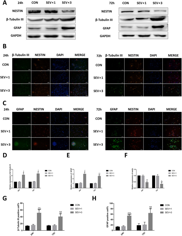
Fig. 3.
Repeated exposure to 4.1% sevoflurane led to early differentiation in primary cultured hippocampal NSCs. a Western blotting images of β-tubulin III, GFAP, and nestin. b Immunofluorescence images of β-tubulin III (green) and nestin (red). Scale bar = 100 μm. c Immunofluorescence images of GFAP (green) and nestin (red). Scale bar = 100 μm. d Quantitative analysis of β-tubulin III. e Quantitative analysis of GFAP. (F) Quantitative analysis of nestin. g Quantification of β-tubulin III-positive cells. h Quantification of GFAP-positive cells. Values are means ± SD (n = 3/group). *p < 0.05, **p < 0.01 compared with CON group; #p < 0.05, ##p < 0.01 compared with SEV × 1 group. One-way ANOVA followed by Tukey’s post hoc multiple comparison test was used for data analysis

