Figure 3.
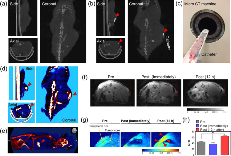
In vivo image-guided injection of the nano-bio therapeutic emulsion into PC3 tumor-bearing mice. (a–b) Micro CT image of the animal model (a) before and (b) after insertion of the catheter in the core region of the tumor. (c) Digital photograph image of the animal model, with a catheter inserted into the tumor site for micro CT imaging. (d) Micro CT and (e) 3D reconstructed the micro CT image of the animal model after the infusion of the nano-bio therapeutic emulsion into the tumor site. (f–g) T1-weighted MR and color-coded MR image of the animal model before and after injection of the nano-bio therapeutic emulsion. (h) Changes in ROI of the tumor site over time in T1-weighted MR images (n=3, *P<0.002).

