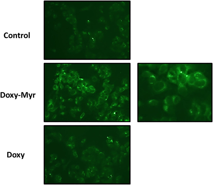
Figure 3.
Doxy-Myr is better retained within cells and reveals a peri-nuclear staining pattern. MCF7 cells were cultured for 72 h as 2D-monolayers, in the presence of Doxycycline or Doxy-Myr, at a concentration of 10 μM. Vehicle-alone controls were processed in parallel. Then, MCF7 cells were washed twice with PBS and subjected to live cell imaging to capture the auto-fluorescent signal retained within cells. Note that Doxy-Myr showed the strongest intracellular retention, and was concentrated within peri-nuclear intracellular compartments. No nuclear staining was observed. Quantitation of mean pixel intensity, using Image J software, revealed that relative to Doxycycline, Doxy-Myr showed a near 3-fold increase in intracellular fluorescence.

