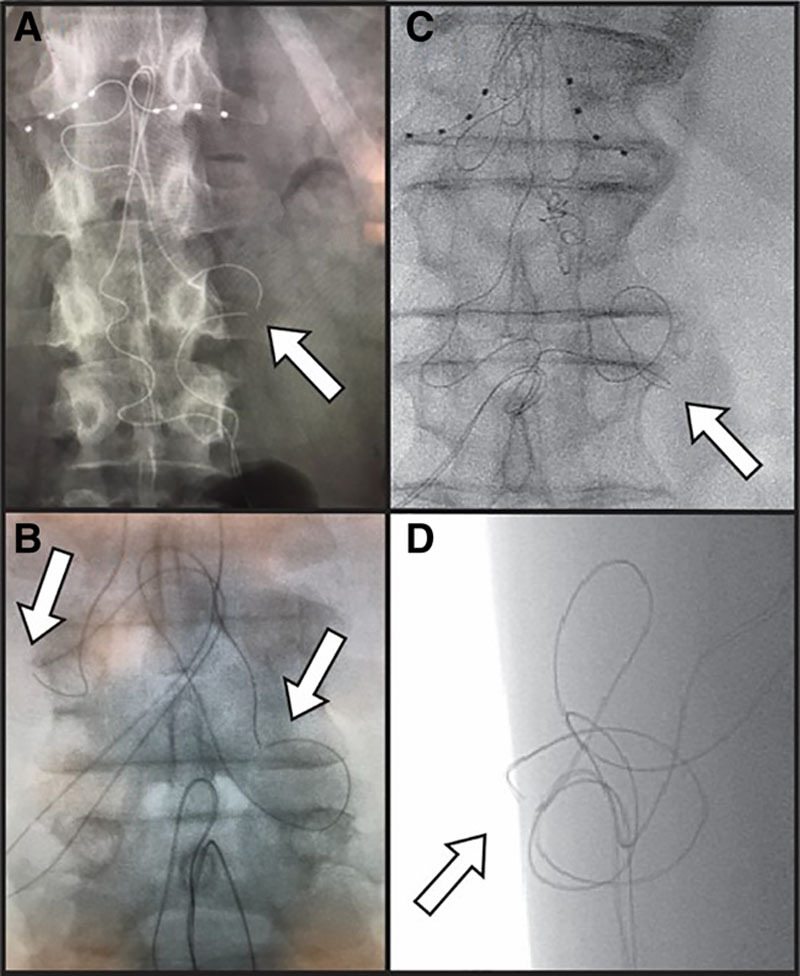
Figure 2.

Visualized lead fractures demonstrated on fluoroscopy. A, A visible lead fracture of a right L1 lead. B, Visible lead fractures at the bilateral L1 DRG-S leads. C and D, Anteroposterior and lateral fluoroscopic views of lead fracture occurring in a superficial plane at the Tuohy needle entry point. The separation of the internal electrical components was visualized in all leads and all leads had intact outer lead sheaths at the time of revision. DRG-S indicates dorsal root ganglion stimulation.
