
Figure 3.
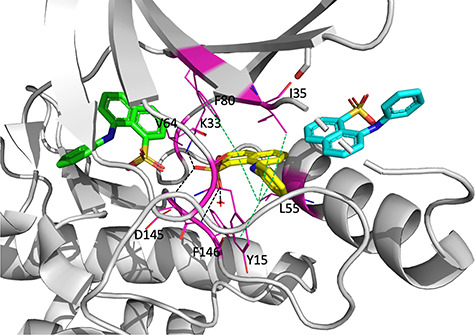
The co-crystal structure of three molecules of ANS bind to CDK2 (PDB ID: 3PXQ) where one of them binds to the ATP-binding site (green), and the other two bind deeper within the allosteric pocket (yellow and cyan). The red cross is a water molecule. Black dotted lines indicate hydrogen bonds and salt bridges. Green dotted lines indicate hydrophobic interactions. Magentas residues interact with the ANS molecule deep within the allosteric pocket (yellow).