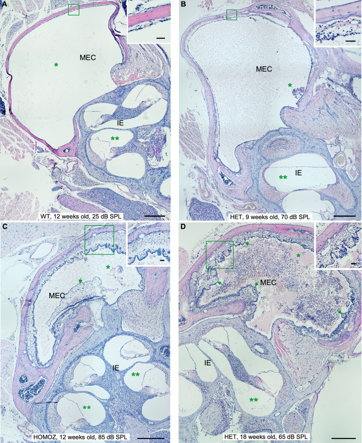
Fig 2. Manifestations of middle ear inflammation in STAT1-KO mice at different ages.
From mild to chronic inflammation degrees in middle ear and cochlea. (A) Wild type (WT) mice at 18 weeks of age, heterozygous (HET) STAT1-KO mice at 9 (B), homozygous (HOMOZ) mice at 12 (C) and heterozygous (HET) mice at 18 (D) weeks of age. Clear appearance of middle ear cavity (MEC) of wild type in comparison to MEC of knockout mice filled with watery effusion with few inflammatory cells (asterisk in B, C) and large number of inflammatory cells (asterisk in D). The cochlea showed no effusion in wild type (two asterisks in A) or effusion with few inflammatory cells in knockout mice (two asterisks in D). The thickness of the middle ear epithelium is greater in knockout mice (insets). Additional signs of inflammation in knockout mice included fibroblastic tissue (arrow in C) and fibrous polyps (arrows in D). Scale bars 300 μm. Inset scale bars 30μm. CO, cochlea; EAC, external auditory canal.

