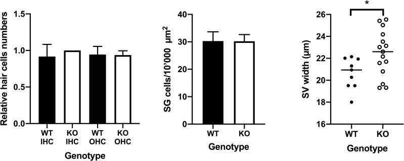
Fig 6. No loss of HC and SG cells in 12–13 weeks old mice.
(A) Comparison of the relative cell numbers of inner hair cells (IHC) and outer hair cells (OHC) between wild type (WT) and STAT1-KO mice (KO). (B) Comparison of spiral ganglion (SG) cells between WT and STAT1-KO mice. (C) Comparison of the stria vascularis (SV) thickness between WT and STAT1-KO mice. STAT1-KO mice show slight thickener membrane as those from the WT mice. Two tailed student’s t-test was used to compare two genotype groups. *P<0.05

