
Figure 4.
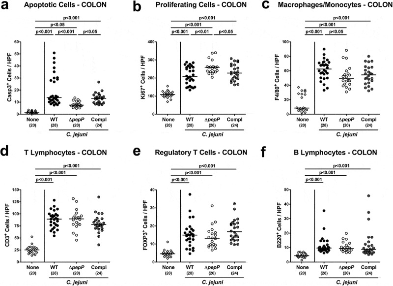
Apoptotic, proliferative/regenerative, and immune cell responses in the colon.
Shown are average numbers obtained from the colon of C.jejuni 81–176 WT (black circles), ΔpepP (white circles) or pepP complemented strain (Compl; crossed circles) infected mice at d6 post-infection for (a) apoptotic cells that are positive for caspase-3, (b) proliferating/regenerating cells positive for Ki67, (c) macrophages and monocytes positive for F4/80, (d) CD3+ Tlymphocytes, (e) FOXP3+ regulatory Tcells, and (f) B220+ Blymphocytes. All data are obtained from six high power fields (400x magnification) per mouse. Uninfected control mice received vehicle only (none, white diamonds). Medians (black bars), significance levels (p-values) determined by the one-sided ANOVA test and Tukey post-correction or the Kruskal–Wallis test and Dunn’s post-correction and the numbers of analyzed mice (in parentheses) are indicated. Data were pooled from four independent experiments.