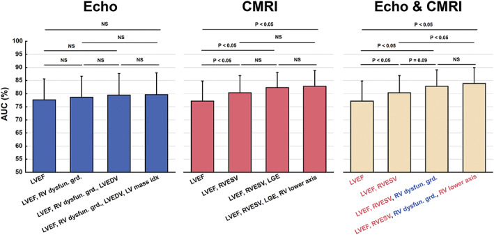
Figure 2.

The results of receiver operating characteristic analyses on the classification outcomes obtained when using only echo or CMRI markers and a combination of both markers. For the latter case, the selected echo and CMRI markers are written in blue and red, respectively. The classification results at different steps of the parameter selection process were statistically compared. AUC, area under the curve; Echo, echocardiogram; CMRI, cardiac magnetic resonance imaging; NS, not significant; LGE, late gadolinium enhancement; LV, left vetricular; LVEDV, left ventricular end‐diastolic volume; LVEF, left ventricular ejection fraction; RV, right ventricular; Dysfun.grd, dysfunction grade; RVESV, right ventricular end‐systolic volume.
