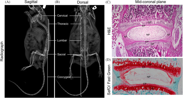
FIGURE 2.

Representative radiograph images in the sagittal, A and dorsal, B plane showing the cervical, thoracic, lumbar, sacral, and coccygeal vertebrae in the spine of a 15‐month‐old male mouse. Representative mid‐coronal section of the lumbar intervertebral disc from a three‐week‐old mouse stained with H&E, C, shows the major components of the disc; SafO/Fast Green, D, staining shows the relative distribution of proteoglycan (stained orange‐red) and collagen (stained fast green). AF, annulus fibrosus; EP, endplate; GP, growth plate; NP, nucleus pulposus
