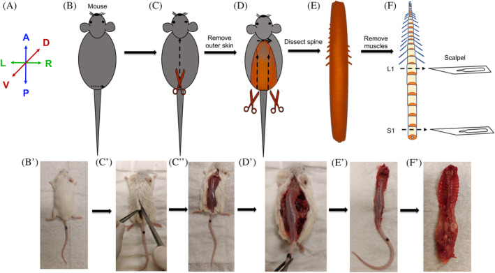
FIGURE 3.

Method for dissection and collection of the mouse spine. A, Anatomical positions for the images presented in B‐F. B and B′, Mouse is placed prone with the ventral side on the dissection table and its back or dorsal side facing the experimenter and doused with 70% ethanol to avoid fur contamination. C, C″, Lift the fur right above the pelvic girdle and create a transverse incision and dissect through the fur from caudal to rostral direction. D and D′, Using a pair of scissors cut from hip joint moving from caudal to rostral end along the lateral side of the spine to dissect the spine. E and E′, Detach and excise the spine from the body. F, Clean the spine by dissecting out the attached muscles. Separate the caudal, lumbar, and thoracic regions of the spine using a scalpel blade or using a pair of fine‐tip and sharp scissors. F′, The ventral view of an excised spine. Images of the mouse were photographed using a cell‐phone camera and are not to scale. A, anterior. P, posterior. R, right. L, left. V, ventral. D, dorsal. L1, first lumbar vertebra. S1, first sacral vertebra
