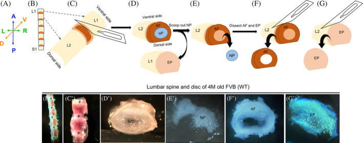
FIGURE 4.

Microdissection procedure for the isolation of the NP, AF, and EP from the intervertebral disc of mouse spine performed using a bright field stereomicroscope. Representative images of the dissected lumbar spine in ventral view, B‐C′, and intervertebral disc in transverse view demonstrating the microdissection procedure, D′,D″, E′, F′ G′, and G″) from a four‐month‐old (4 M) WT FVB mouse captured using an iPhone camera and bright field stereomicroscope. A, Anatomical positions for the images shown in B‐G′. Place the dissected lumbar spine with the dorsal side on the Petri Dish and ventral side facing the experimenter, B and B′. Dissect at the AF and EP junction, C‐C″ to expose gelatinous NP surrounded by AF, D. Transverse view of an intact IVD (D′). Scoop out NP from the AF (E and E′) and collect. Microdissect the AF, F, and F′, and collect. Microdissect the EP, G and G′ and collect. A, anterior. P, posterior. R, right. L, left. V, ventral. D, dorsal. IVD, intervertebral disc. NP, nucleus pulposus. AF, annulus fibrosus. EP, end plate. # represents the disc in B′ and C′
