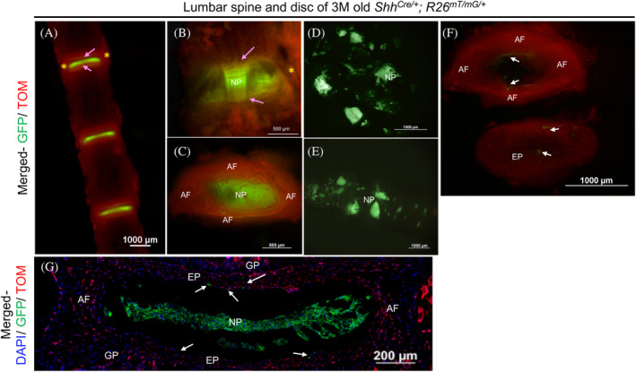
FIGURE 5.

Validation of microdissection procedure using ShhCre/+; R26mT/mG/+ fluorescence reporter line. Representative images of the lumbar spine and intervertebral disc dissected from a three‐month‐old (3 M) ShhCre/+; R26mT/mG/+ mouse where all NP cells appear GFP+ve under an epi‐fluorescence stereomicroscope (A‐G). L1‐L4 spine in ventral view (A), an individual disc at higher magnification in ventral view (B), and a dissected disc in transverse view (C) showing GFP+ve NP cells, TOM+ve AF (yellow asterisk), EP (indicated by pink arrows), and vertebrae from 3 M old ShhCre/+; R26mT/mG/+. NP cells scooped out of the AF (D), collected with a pipette tip (E) are free of any TOM + ve cells. AF and EP are dissected using a fresh scalpel blade (F). C and F are images of the same disc captured in transverse plane. While C shows the intact disc with NP in the center viewed under the thin layer of EP, together surrounded by lamella of AF, F shows the isolated AF and EP following removal of the NP cells. Representative fluorescence image of a mid‐coronal section of a 3 M old ShhCre/+; R26mT/mG/+ with DAPI counterstained nuclei (blue) shows that all notochord‐descendant NP cells are GFP+ve (G). A few notochord‐descendant GFP+ve cells are observed in the AF and EP (white arrows in F and G), which may occur during development as previously reported. NP, nucleus pulposus. AF, annulus fibrosus. EP, end plate. * indicates AF on the lateral side of GFP+ve NP cells in A and B. The pink arrow identifies the EP in A and B
