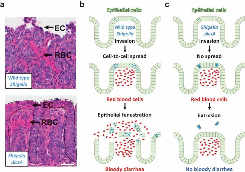
Figure 2.

Connecting Cell Biology and pathogenesis. (a) Representative images of hematoxylin- and eosin-stained colonic mucosa of infant rabbit colon infected with wild type S. flexneri (top) and the ΔicsA mutant (bottom). EC, epithelial cell; RBC red blood cell. Dotted lines delineate crypts. Scale bar, 50 μm. (b) Top. Wild type S. flexneri (blue) invades the colonic epithelium (green). Middle. Wild type bacteria spread from cell to cell. Intracellular infection leads to vascular lesions and red blood cell infiltration (red). Bottom. The combination of epithelial fenestration due to cell-to-cell spread and red blood cell infiltration leads to bloody diarrhea. (c) Top. The ΔicsA mutant (blue) invades the colonic epithelium (green). Middle. The ΔicsA mutant does not spread from cell to cell and grows as macro-colonies. Intracellular infection leads to vascular lesions and red blood cell infiltration (red). Bottom. In absence of cell-to-cell spread, cells infected with the ΔicsA mutant are eliminated, presumably through extrusion. The epithelial structure remains intact, and the animals do not experience bloody diarrhea, or any signs of illness.
