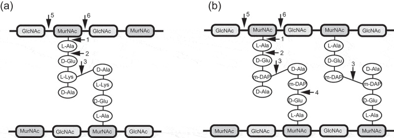
Figure 2.

Schemetic presentation of bacterial peptidoglycan and generalized cut sites of peptidoglycan hydrolases. (a) A3α type peptidoglycan of Staphylococcus aureus; (b) A1γ type featuring peptidoglycan of C. difficile. The bonds potentially attacked by endolysins of different enzymatic specificities are indicated by numbers: 1) N-acetylmuramoyl-L-alanine amidases; 2) L-alanoyl-D-glutamate endopeptidases; 3) interpeptide bridge endopeptidases; 4) D-glutamyl-m-DAP endopeptidase; 5) N-acetyl-D-glucosaminidases; 6) N-acetyl-D-muramidases and lytic transglycosylases.
