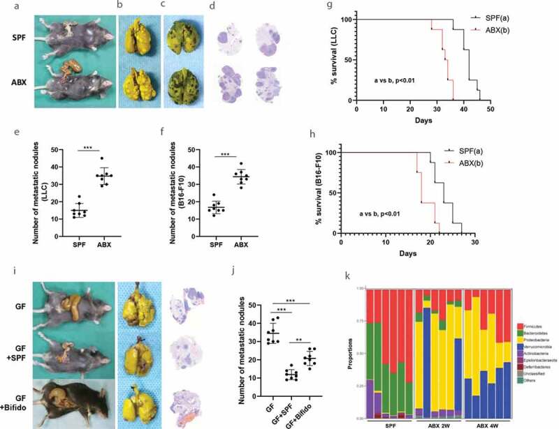
Figure 1.

Gut microbiota dysbiosis promotes metastasis in animal experiment model. A-F, Tumor mice models were established by inoculating with LLC cells (b) or melanoma B16-F10 cells (c) via tail vein into C57BL/6 mice. The visible differences in the number of metastatic nodules between SPF and SPF/ABX mice were observed as a whole lung (b-c) or by pathological and HE stain (d). The number of metastatic nodules between different groups was calculated (e-f). G-H, Survival curves analysis. Kaplan-Meier estimates for survival rate of the SPF and SPF/ABX-treated mice. p values are shown [log-rank (Mantel-Cox) test analysis]. I-J, The visible differences in the number of metastatic nodules among GF, GF/SPF or GF/Bifido group were analyzed as a whole lung or by HE staining. These experiments were performed in two sets and 6–8 mice per group. K, By 16 S rDNA sequencing, mice fecal samples were sequenced to evaluate the influence of antibiotics on gut microbiota in different groups. Statistical analysis: one-way ANOVA (Figure 1E-F,1 J). Data are shown as mean ± SEM. *P < .05; **P < .01; ***P < .001.
