
Figure 5.
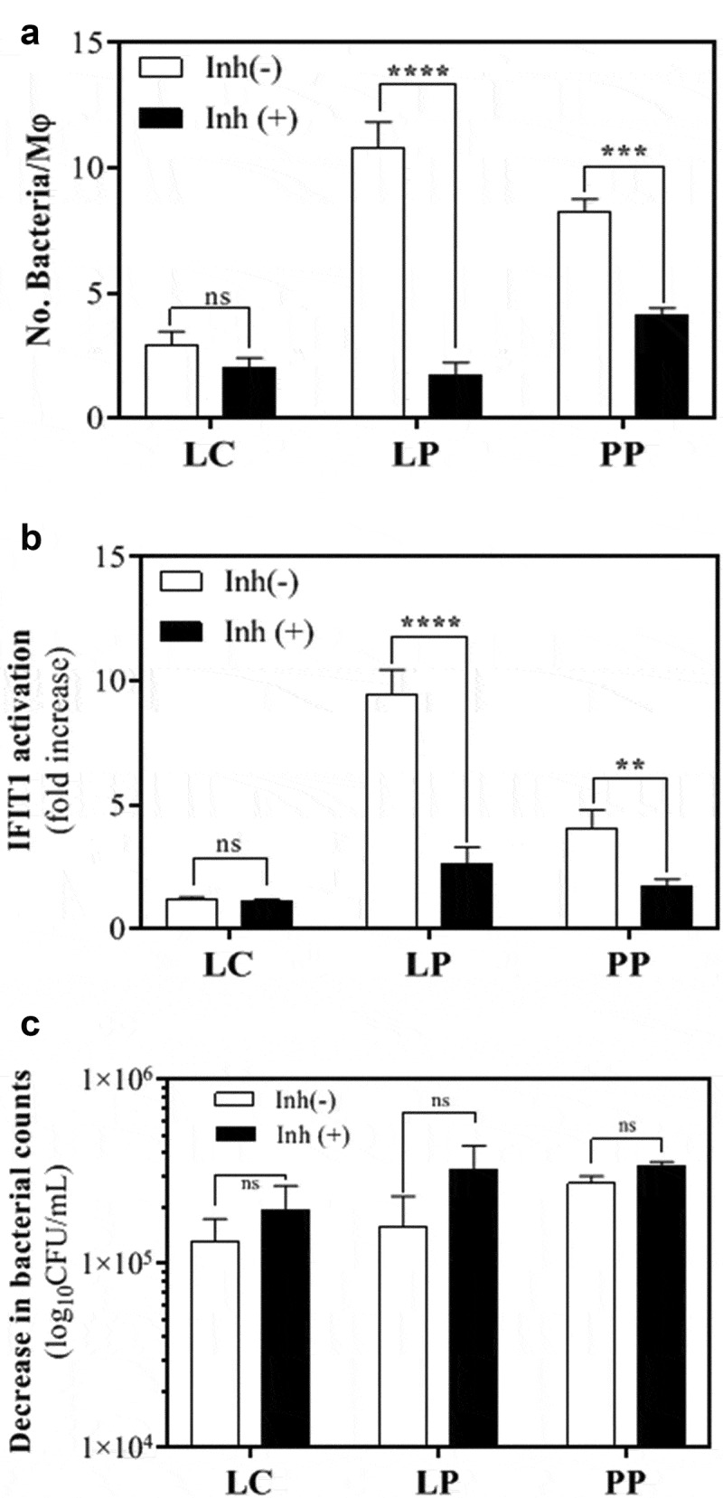
Human THP-1 macrophages interact with Lactobacillus plantarum (LP) and Pediococcus pentosaceus (PP). (a) Number of L. casei (LC), LP or PP cells that were internalized and/or attached to THP1 macrophages after a 2 h RPMI incubation in the absence (white) or presence (black) of the phagocytosis inhibitor cytochalasin D. The bacterial intake and/or attachment was calculated as the difference between the number of bacteria found in the supernatant of THP-1 cells at 0 and 2 h post-LAB challenge, and presented as number of bacteria per macrophage (Mφ). Comparative analysis was carried out using the Student t-test (ns, no significant; ***p < .005; ****p < .001). (b) IFIT1 activation in PMA-differentiated IFIT1-GLuc THP-1 macrophages exposed to LC, LP or PP in the absence (white) or presence (black) of cytochalasin D. The activation of IFIT1 is presented as a fold increase over a non-stimulated condition. The bacterial challenge was carried out at a ratio of 1 macrophage per 25 bacteria (**p < .01; ****p < .001; Student’s t-test). (c) Decrease in bacterial counts for LC, LP and PP after a 2 h RPMI incubation in the absence (white) or presence (black) of cytochalasin D. The bacterial decrease was calculated as the difference between the number of viable bacteria after 2 h of incubation and expressed as log10CFU/mL (ns, no significant; ***p < .005, ****p < .001; Student’s t-test).