
Figure 1.
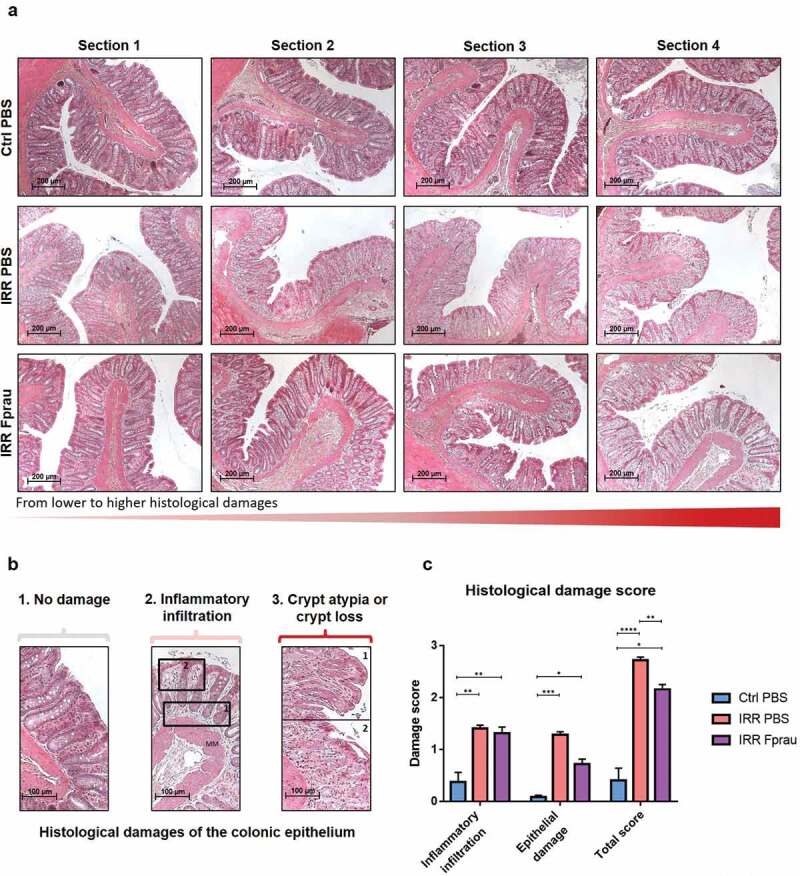
Effect of prophylactic F. prausnitzii treatment on 29 Gy colorectal irradiation-induced histological damage to the colonic mucosa at 3 d.
(a-b) Colonic sections were stained with hematoxylin, eosin and saffron (HES). (a) Sections show histological structure change of the colonic mucosa in one representative rat (4 representative sections/rat) of each group, the control group, the irradiated group and the irradiated and F. prausnitzii-treated group (Scale bars 200 µm). (b) Representative pictures showing parameters considered for histological scoring. (b1) No damage, (b2) inflammatory infiltration with a score graduation between 0 and 1 for modest inflammatory infiltration leading to slight crypt detachment from the muscularis mucosa (MM, rectangle 1) or between 1 and 2 for inflammatory infiltration observed in all the lamina propria area (rectangle 2) and (b3) epithelial damages with a score graduation between 0 and 1 for crypt morphological atypia characterized by disorganized and deformed epithelial crypts (rectangle 1) or between 1 and 2 for crypt loss (rectangle 2). Scale bars 100 µm (c) Histogram represents means±SEM of inflammatory infiltration score, epithelial damage score or total scoring including inflammatory infiltration score and epithelial damage score (for total scoring graduation of the injury was: score 0 = null; 0< score<1 = slight; 1< score<2 = moderate and score>2 = severe) quantified in control animals, in irradiated animals or in irradiated and F. prausnitzii-treated animals. Analyses were performed on 20 sections per animal and 5 animals were used per group (N = 1). Ctrl = Control, IRR = Irradiated, Fprau = F. prausnitzii. Error bars represent SEM, *p <.05, **p <.01, ***p <.001, ****p <.0001.