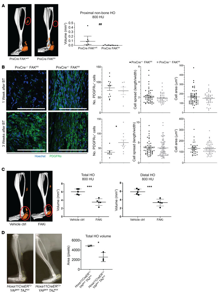
Figure 3. FAK deletion or inhibition reduces heterotopic bone.
(A) Deletion of FAK (Ptk2) within the Prx lineage reduces proximal non–bone HO compared with mouse with single wt allele in Prx lineage by μCT imaging at 800 Hounsfield units (HU) (##P < 0.01, n = 6–9/group, Mann-Whitney U test). (B) Confocal microscopy images of Prx-Cre– and Prx-Cre+ deletion of FAK probed with indicated antibodies at 1 and 3 week time points after injury (left) quantified PDGFRα cell number, cell spread, and cell area (*P < 0.05, n = 2–4/group, n = 2–4 roi/mouse, n > 15 cells/image, Student’s t test). Scale bars: 100 µm. (C) FAK inhibitor (FAKi) PF573228 treated mice showed reduced total and distal HO at 800 HU 9 weeks after injury (***P < 0.001, n = 5/group, Student’s t test). (D) Inducible conditional deletion of YAP and TAZ coactivators within Hoxa11-expressing cells causes 50% reduction in ectopic bone formation Student’s t test (*P < 0.05, n = 2–3 mice/group).

