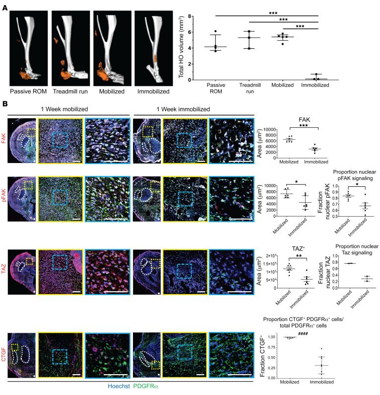
Figure 4. Hind limb immobilization reduced HO formation and alters cell fate.
(A) μCT imaging of passive range of motion, forced mobilization, normal ambulation, and complete immobilization groups 9 weeks after injury with reconstructions of representative means at 800 HU show reduced HO formation in immobilized hind limb (***P < 0.001, n = 3–4/group). (B) Confocal microscopy images of injured hind limb cross sections with indicated antibody probes at 1 week after injury with quantifications of FAK, pFAK, and TAZ (right) (n = 3/group, n = 1–3 roi/mouse, *P < 0.05, **P < 0.01, ***P < 0.001) (####P < 0.0001, Mann-Whitney U test). Scale bars: 100 µm. Calculated using ANOVA (A) or Student’s t test (B).

