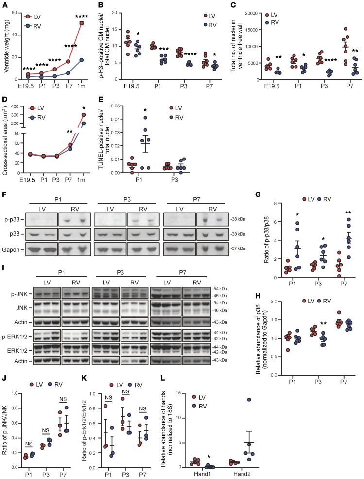
Figure 1. Chamber-specific remodeling and activation of p38 MAPK during postnatal heart development.
(A) The tissue weights of the left (LV) and the right (RV) ventricles of mouse perinatal heart at different time points as indicated (E19.5, n = 12; P1, n = 16; P3, n = 11 ; P7, n = 9; 1 month, n = 9). (B) The phospho–histone H3–positive (p-H3–positive) cardiomyocyte (CM) nuclei/total CM nuclei ratio in the LV vs. RV (E19.5 and P1, n = 6; P3 and P7, n = 7). (C) Total number of CM nuclei (E19.5 and P1, n = 6; P3 and P7, n = 7). (D) The cross-sectional area of CMs (E19.5 and P7, n = 6; 1 month, n = 3). (E) The number of TUNEL-positive CMs (n = 6). (F) Representative immunoblots of phospho-p38, total p38, and Gapdh. (G and H) The phospho-p38 vs. total p38 signal ratio (G) and total p38 (H) (n = 6). (I) Representative immunoblots of phosphorylated ERK and JNK vs. total ERK and total JNK in mouse ventricles. (J and K) The phosphorylated JNK vs. total JNK signal ratio (J) and phosphorylated ERK vs. total ERK signal ratio (K) (n = 3). (L) Chamber specificity of mRNA expression of Hand1 vs. Hand2 detected in the LV vs. RV free wall prepared from P7 neonatal hearts (n = 5). For all panels, data are presented as mean ± SEM. ****P < 0.0001, ***P < 0.001, **P < 0.01, *P < 0.05 (RV vs. LV). See complete unedited blots in the supplemental material.

