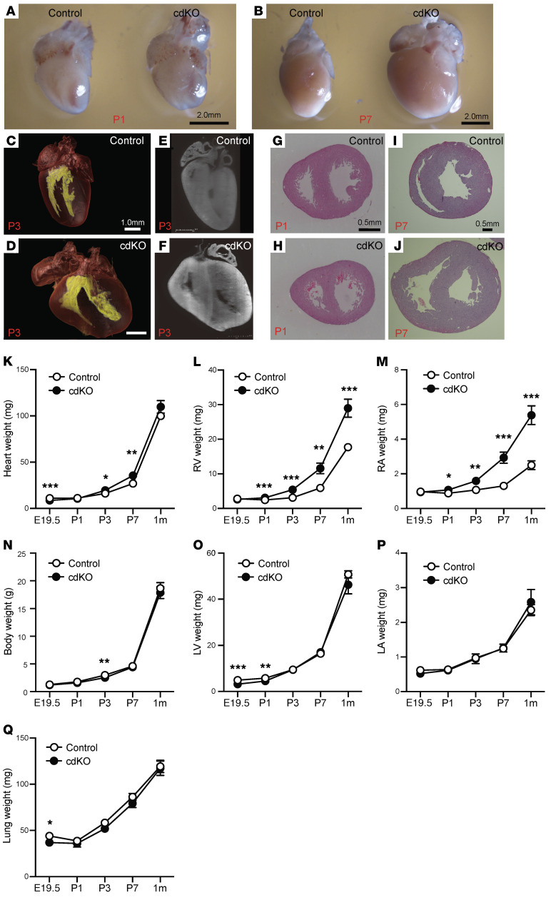
Figure 2. RV-specific abnormality in the cardiomyocyte-specific p38α/β MAPK–knockout mouse.
(A and B) Whole-heart images of control and p38-cdKO at P1 (A) and P7 (B). (C–F) Light-sheet imaging of whole heart from control (C) and p38-cdKO (D) at P3. Inner cavity of ventricles is labeled in yellow. Sliced image from light-sheet imaging from control (E) and p38-cdKO (F) at P3. (G and H) H&E-stained histological section of control heart (G) and p38-cdKO heart (H) at P1. (I and J) H&E-stained histological section of control heart (I) and p38-cdKO heart (J) at P7. (K–Q) Weight measurements of whole heart (K), RV (L), right atrium (RA) (M), whole body (N), LV (O), left atrium (LA) (P), and lung (Q) in control and p38-cdKO mice during neonatal development (n = 12 for E19.5, n = 16 for P1, n = 11 for P3, n = 9 for P7, n = 9 for 1 month; mean ± SEM). ***P < 0.001, **P < 0.01, *P < 0.05 for all panels (control vs. p38-cdKO).

