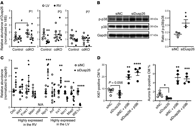
Figure 7. DUSP26 expression upon chamber-specific p38 activation.
(A) Chamber-specific DUSP26 mRNA levels in the neonatal mouse heart at P1, P3, and P7 as indicated (n = 6; mean ± SEM). **P < 0.01, *P < 0.05 (LV vs. RV); #P < 0.05 (control vs. p38-cdKO). (B) p38 activation in cardiomyocytes treated with siRNA against Dusp26 (n = 3; mean ± SEM). (C) mRNA expression of genes differentially expressed in each ventricle in the siDusp26-treated cardiomyocytes (n = 6; mean ± SEM). (D and E) The levels of Ki67-positive (D) or Aurora B–positive (E) cardiomyocytes (CM) following different treatments with p38 inhibitor (p38i, SB202190) and siDusp26 (n = 4; mean ± SEM). ****P < 0.0001, ***P < 0.001, **P < 0.01, *P < 0.05 (vs. scrambled siRNA control [siNC]).

